R&D Focus
ITRI’s Innovations in E-Health Monitoring and Inspection
ITRI presented five e-health monitoring and inspection technologies including In-Sleep Target Memory Consolidation Technology, the Handheld Skin Quality Optical Coherence Tomography system, the Handheld Ultrasound System, the Measurement Technique for Lumbar Spine X-Ray Images and the AI Decision Support Technology of Fundus Image in Diabetes Mellitus at this year’s CES digital show. These innovations highlight the importance of e-health technology that can provide efficient and accurate detection and examination, helping individuals maintain wellness and achieve better quality of life.
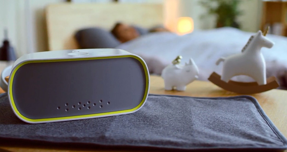
In-Sleep Target Memory Consolidation (ISTMC) Technology strengthens memory of selected subjects.
In-Sleep Target Memory Consolidation (ISTMC) Technology
ITRI’s In-Sleep Target Memory Consolidation (ISTMC) Technology offers users a breakthrough capability of seamlessly strengthening the memorization of selective subjects without wearing any sensors such as electroencephalography (EEG) sensors. During the day, a user reads out loud the subjects to be memorized, such as new words and phrases of a foreign language, anatomical details of the human body, provisions of an election law, etc., and ITRI’s ISTMC device records them. During the night, ITRI’s ISTMC device detects when the user is in the deep sleep stage and replays the recorded sounds to reinforce the brain’s process of transferring the sounds’ corresponding memorized subjects from short-term memory to long-term memory. The key enabling technology of this innovative device is the ability to detect, reliably and accurately, when a user enters the deep sleep stage based solely on the user’s breathing rate, heart rate and body movement, which in turn are measured by the application of recurrent neural network technology to the channel state information (CSI) in the Wi-Fi signals emitted by the user’s smartphone.
Video of ISTMC.
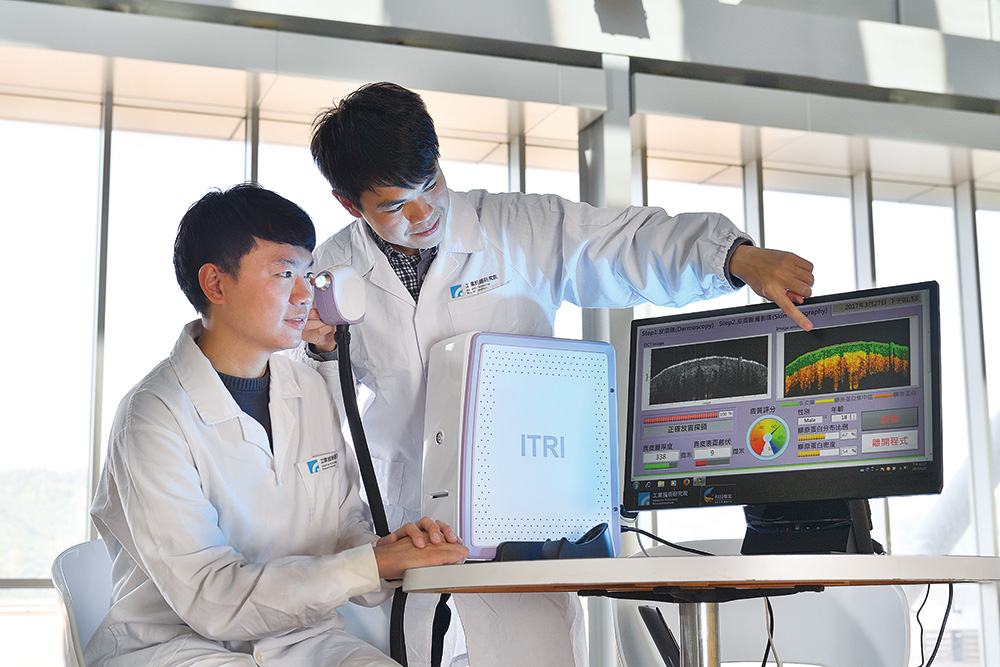
Handheld Skin Quality Optical Coherence Tomography
ITRI developed Handheld Skin Quality Optical Coherence Tomography (OCT), the first handheld OCT system that analyzes subsurface skin structures and detects collagen distribution in the dermis layer. The system replaces the conventional approach of an invasive biopsy for examining tissue underneath the skin. It incorporates a built-in skin quality analysis model, AI, and anti-shake image capturing technology to evaluate skin quality within 10 seconds. It weighs less than 400 grams (14 ounces), allowing users to operate the device with one hand. This innovation is used for medical purposes and provides valid scientific proof for product development in the cosmetic industry, shortening time-to-market while reducing the number of animals used in experiments. The device combines an optomechanical-electronics controller and an image processor to provide 3D optical biopsy using a broadband near-infrared light source. This user-friendly system features high resolution (of few microns) and a fast scan rate. It determines skin quality by analyzing key parameters including dermis and epidermis thickness, collagen density, and pore size as well as the number of blood vessels.
Video of Handheld Skin Quality Optical Coherence Tomography.
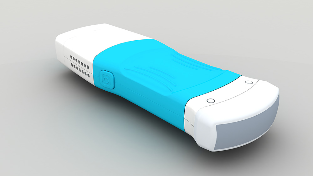
Handheld Ultrasound System
The Handheld Ultrasound System enables healthcare professionals to perform ultrasound exams anywhere, anytime. It saves critical time in emergency and intensive care. The system includes a lightweight, portable scanner and a mobile application. The 64-channel system supports changeable transducers (linear 7.5MHz/convex 3.5MHz) and can process B-mode and color Doppler mode images. In the future, deep learning technology can be integrated to perform quantitative analysis for diagnosis of liver diseases.
Video of Handheld Ultrasound System.
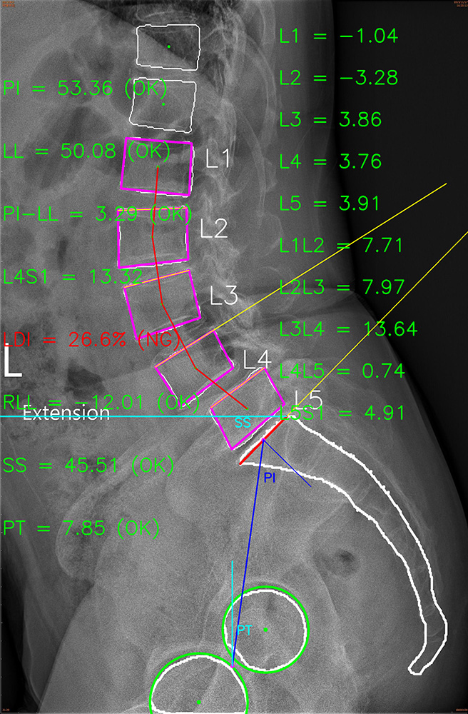
Measurement Technique for Lumbar Spine X-Ray Images
The Measurement Technique for Lumbar Spine X-Ray Images combines deep learning and image processing algorithms to reduce time and labor for lumbar spine X-ray interpretation. The system provides real-time measurements of pelvic incidence (PI) and lumbar lordosis (LL), and abnormal findings such as spondylolisthesis. It also can assist in surgical planning and offers precise calculation of surgical implants for clinical diagnosis. The system uses AI to conduct X-ray imaging simulation and enable prediction of postoperative PI and LL values for clinical and auxiliary analysis.
Video of Measurement Technique for Lumbar Spine X-Ray Images.
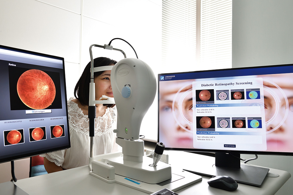
AI Decision Support Technology of Fundus Image in Diabetes Mellitus
The AI Decision Support Technology of Fundus Image in Diabetes Mellitus is the first AI technology to enable early detection and timely treatment of diabetic retinopathy (DR) by classifying and locating lesions and determining DR severity. It locates the four main DR lesions including microaneurysms, hemorrhages, soft exudates, and hard exudates, and classifies examination results into one of the five stages of DR (no DR; mild, moderate, or severe non-proliferative DR; or proliferative DR). The technology uses AI to provide binary decision support for ophthalmologist referrals, and optimal treatment timing for Clinical Significant Macular Edema (CSME).
Video of AI Decision Support Technology of Fundus Image in Diabetes Mellitus.